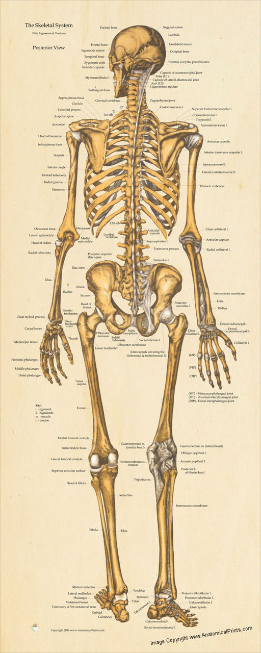
Skeletal System Posterior View Poster Clinical Charts and Supplies
posterior view. previous. next. gastrocnemius Large thick muscle forming the curve of the calf and allowing the foot to extend; it also helps the knee to extend. gracilis Muscle enabling the thigh to draw near the median axis of the body, and the leg to flex on the thigh and to rotate inwardly (toward the median axis). biceps of thigh.
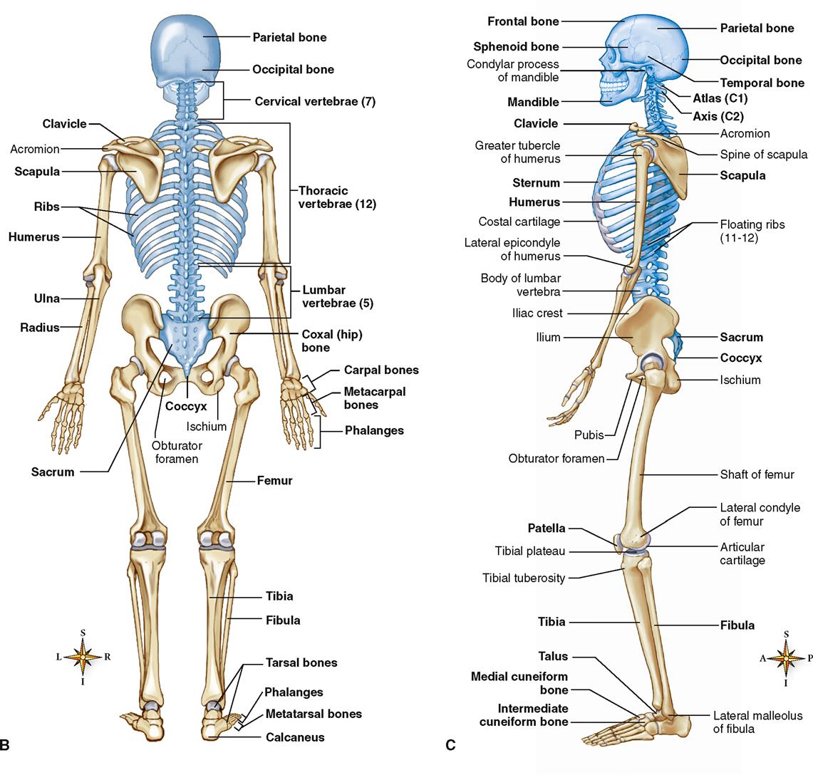
Skeletal System Basicmedical Key
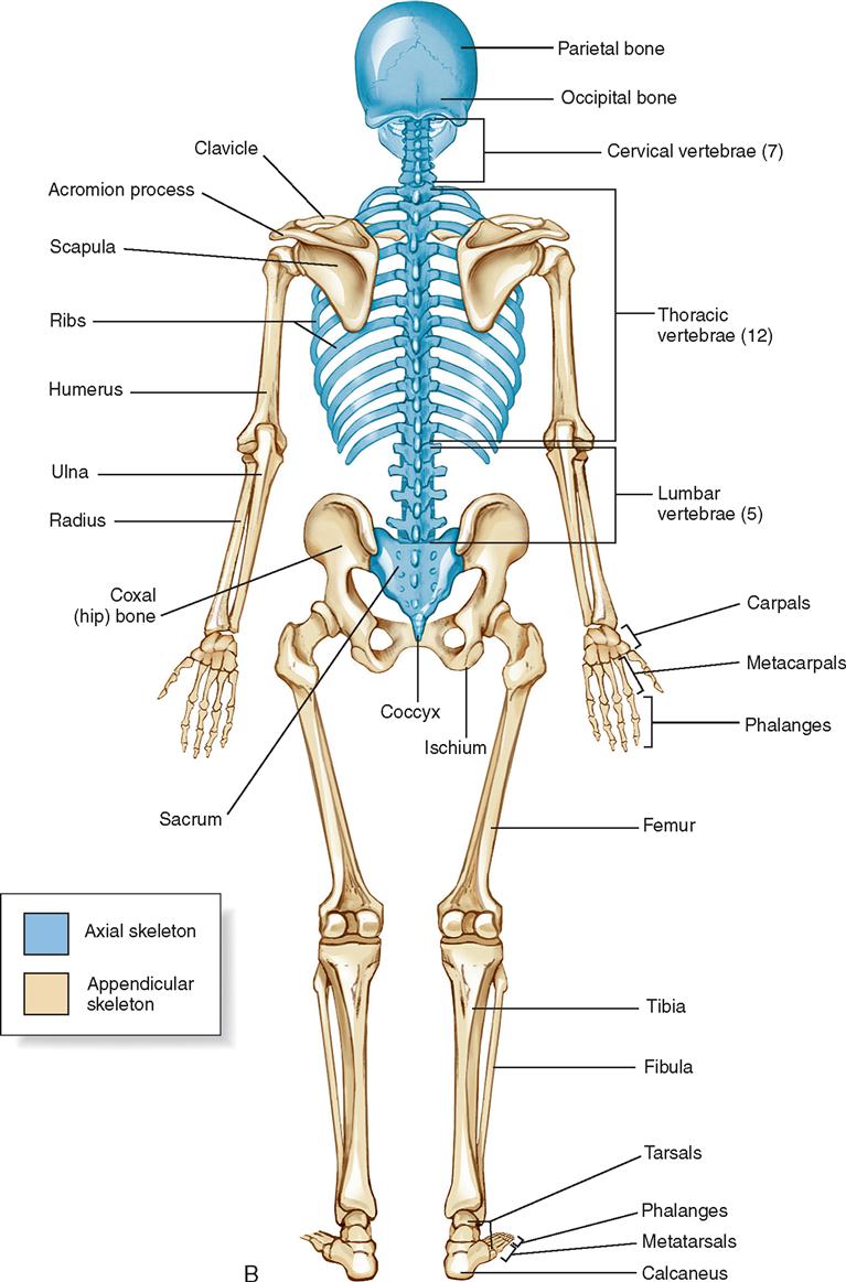
Skeleton—Posterior View Bones of the Skull—Frontal View Bones of the Skull—Lateral View Types of Fractures Types of Traction Types of Synovial Joints For an in-depth study of the skeletal system, consult the following publications: Lewis SM, et al: Medical-surgical nursing, ed 8, St. Louis, 2011, Mosby.

Illustration of anterior and posterior views of human skeletal
Dec. 24, 2023, 4:25 AM ET (Yahoo News) Human skeletons, remains of sharks, blood-sucking bats. human skeleton, the internal skeleton that serves as a framework for the body. This framework consists of many individual bones and cartilages.
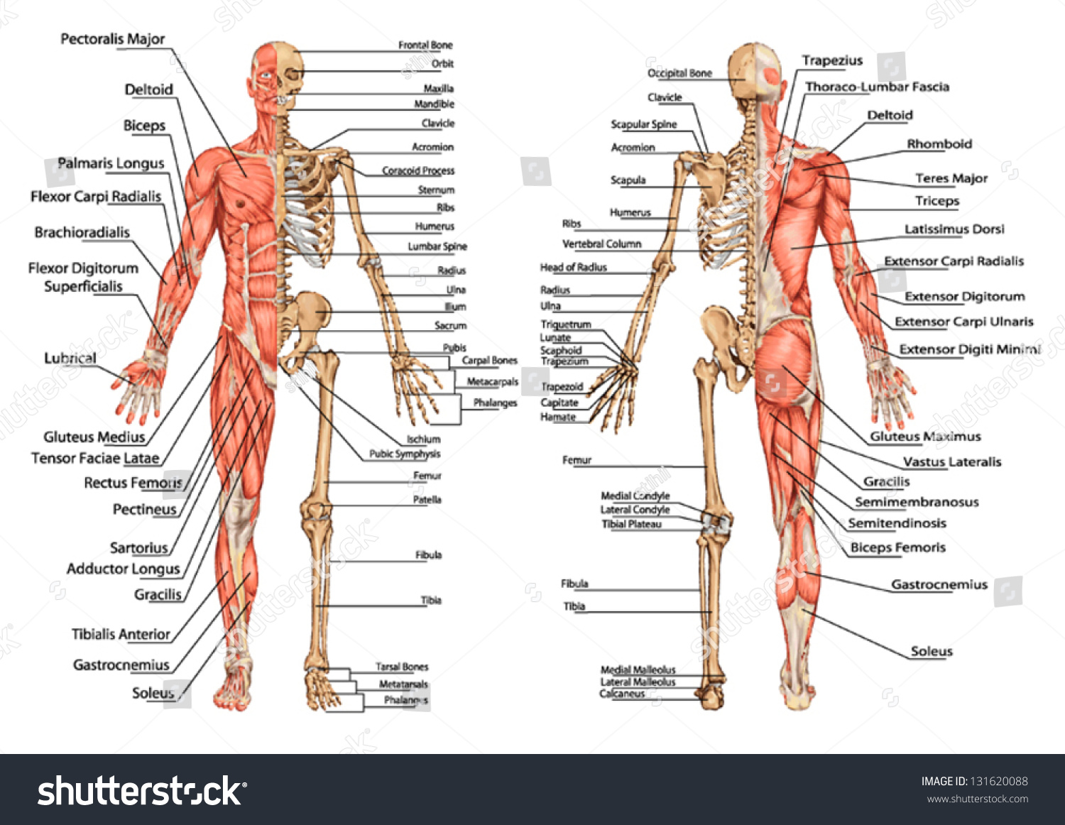
lateral human anatomy
The Skull Bones Anatomy - Inferior View. A number of cranial and facial bones are visible when viewing the skull inferiorly. Review the bones of the skull and test your knowledge. The Skull Bones - Orbital View. There are a number of markings on the cranial and facial bones which form the orbit of the skull.

Anterior and Posterior view Human bones anatomy, Body bones, Skeleton
Browse 3,100+ posterior view of skeleton stock photos and images available, or start a new search to explore more stock photos and images. Sort by: Most popular. The skeletal system. The human skeletal system, vector illustrations of human skeleton front and rear view. Targeting back pain.
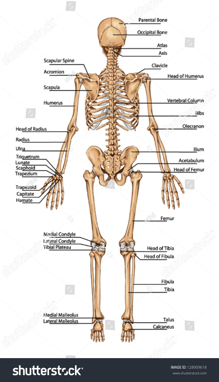
Human Skeleton From The Posterior View Didactic Board Of Anatomy Of
Create healthcare diagrams like this example called View of the Full Skeleton - Posterior in minutes with SmartDraw. SmartDraw includes 1000s of professional healthcare and anatomy chart templates that you can modify and make your own. 3/37 EXAMPLES. EDIT THIS EXAMPLE.
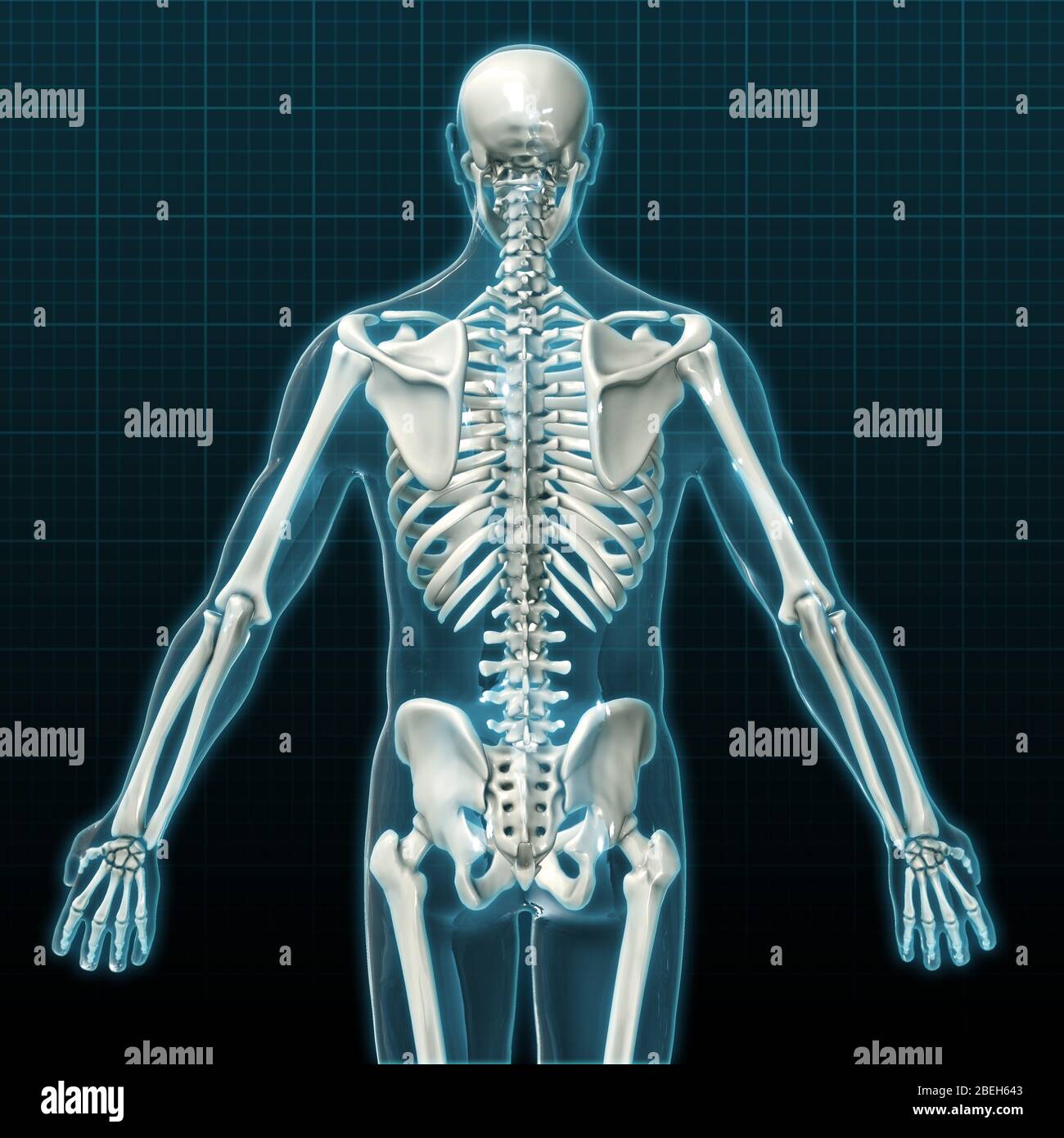
Human skeleton posterior view hires stock photography and images Alamy
HOME :: HUMAN BEING :: ANATOMY :: SKELETON :: POSTERIOR VIEW posterior view previous next parietal bone Flat cranial bone articulating with the frontal, occipital, temporal and sphenoid bones; the two parietal bones form the largest portion of the dome of the skull. lateral view of skull axis
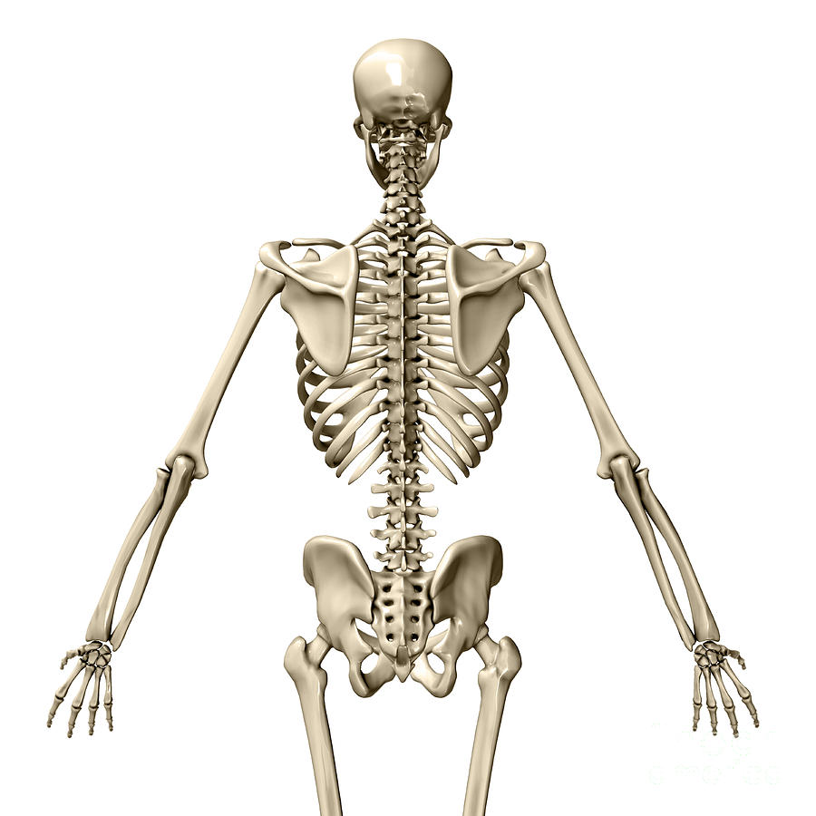
Human Skeleton, Posterior View Photograph by Evan Oto Fine Art America
1/6 Synonyms: Spine The vertebral column (spine or backbone) is a curved structure composed of bony vertebrae that are interconnected by cartilaginous intervertebral discs. It is part of the axial skeleton and extends from the base of the skull to the tip of the coccyx. The spinal cord runs through its center.
Posterior Anterior View
Figure 7.3.2 - Anterior View of Skull: An anterior view of the skull shows the bones that form the forehead, orbits (eye sockets), nasal cavity, nasal septum, and upper and lower jaws. Inside the nasal area of the skull, the nasal cavity is divided into halves by the nasal septum.
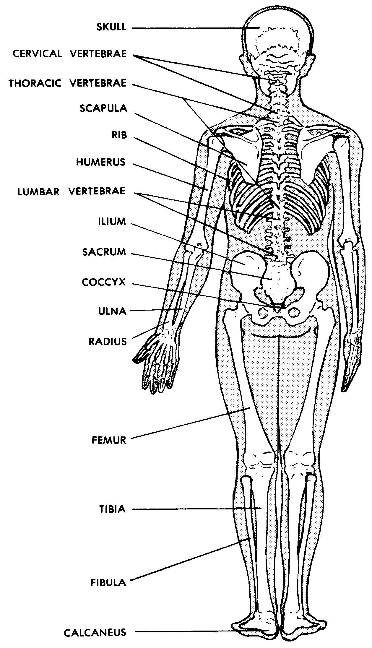
Images 04. Skeletal System Basic Human Anatomy
Skull 3 Lateral - Short - Medium - Text - Answers. Skull 4 Lateral - Short - Medium - Text - Answers. Skull 5 Lateral - Short - Medium - Text - Answers. Skull 6 Lateral - Short - Medium - Text - Answers. Skull 7 Lateral - Short - Medium - Text - Answers. Skull 1 Cranial - Short - Text - Answers. Skull 1 Inferior - Short - Medium - Long - Text.
.PNG)
Skeletal System Presentation Biology
The posterior view of the skeleton reveals bones that are obscured in the anterior view, most notably, the entire stack of individual vertebrae that span vertically from the sacrum to the skull.
upper skeletal anatomy
1/20 Synonyms: none The posterior and lateral views of the skull show us important bones that maintain the integrity of the skull. The posterior surface protects the region of the brain that contains the occipital lobes and cerebellum .
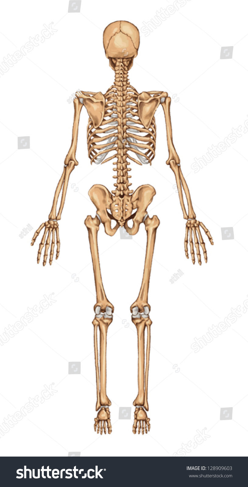
Vektor Stok Human Skeleton Posterior View Didactic Board (Tanpa Royalti
6.1 Skeleton: Overview (See page(s) 84) Name at least five functions of the skeleton. Explain a classification of bones based on their shapes. Describe the anatomy of a long bone. Describe the growth and development of bones. Name and describe six types of fractures, and state the four steps in fracture repair. 6.2 Axial Skeleton (See page(s) 89)
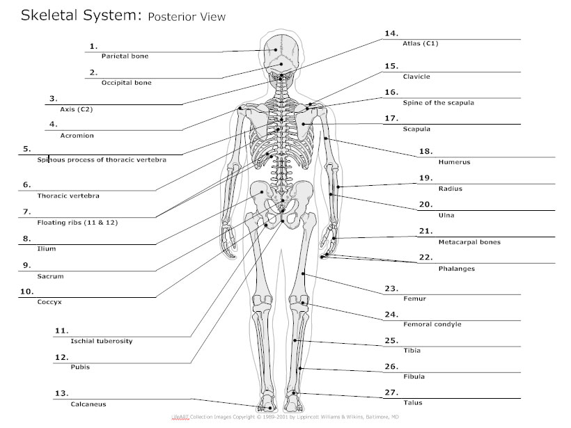
Skeletal System Diagram Types of Skeletal System Diagrams, Examples, More
Lesser Trochanter Obturator Membrane Pelvis Posterior Sacroiliac Ligament Pubic Symphysis Pubofemoral Ligament Sacroiliac Joint Sacrospinous Ligament Sacrotuberous Ligament Sacrum Supraspinous Ligament Change Current View Angle Bones of the Pelvis and Lower Back (Posterior View) Toggle Anatomy System Cardiovascular System of the Lower Torso

View of the Full Skeleton Posterior
This is the midline. Medial means towards the midline, lateral means away from the midline. The eye is lateral to the nose. The nose is medial to the ears. The brachial artery lies medial to the biceps tendon. Fig 1.0 - Anatomical terms of location labelled on the anatomical position.
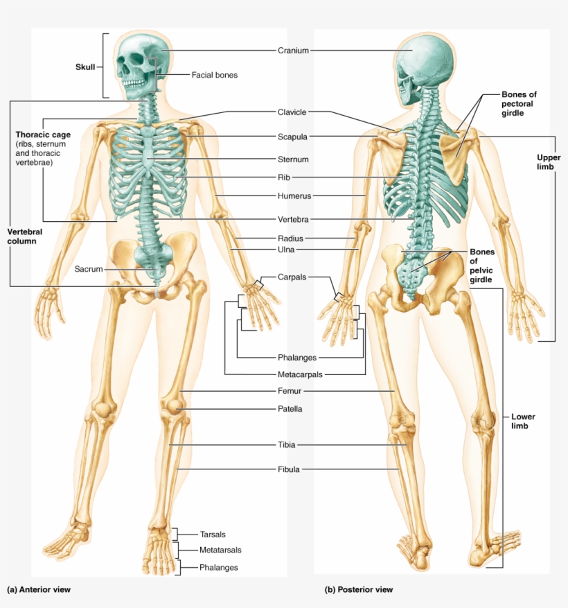
Bones, Part Human Skeleton Anterior And Posterior View PNG Image
Figure 1.4.2 - Directional Terms Applied to the Human Body: Paired directional terms are shown as applied to the human body. is a two-dimensional surface of a three-dimensional structure that has been cut. Modern medical imaging devices enable clinicians to obtain "virtual sections" of living bodies. We call these scans.